Diagnostic methods for the detection of acute gastroenteritis


Diagnostic methods for the detection of acute gastroenteritis

Acute gastroenteritis is among the most common causes of morbidity and mortality worldwide1, with the highest prevalence observed in non-industrialised countries as the disease is most frequently transmitted under conditions of poor hygiene or by consuming contaminated food or water.2 However, acute gastroenteritis remains a common condition in both low- and high-income countries.3 Symptoms and complications have led to a significant impact on workload and economic burden for the healthcare system and society.3 For example, in the Netherlands and Belgium, annual direct medical expenditure due to acute gastroenteritis amounts to 945 million euros and 112 million euros, respectively.3,4 Similarly, in Finland, the loss of efficiency and the associated cost of lost work days were estimated at 1.8-2.1 million euros, with the prevalence of workers absent due to illness recorded at 3.54 times higher in the week following an outbreak.5


Acute gastroenteritis is responsible for 1.45 million deaths worldwide

Gastroenteritis is a transient disease that can be caused by a multitude of different viral, bacterial, and parasitic pathogens.6 Norovirus is the pathogen most commonly responsible for acute gastroenteritis in the United States, followed by Salmonella spp., Shigella spp. and Shiga toxin-producing E-coli (STEC). Among children in particular, Rotavirus is the pathogen of greatest clinical relevance7. It is estimated that, in 2013, Rotavirus was responsible for the deaths of more than 200,000 children worldwide, and the most recent estimates of mortality from this virus range from 500 to 600 deaths per day.7

Improvements in sanitation have led to an overall reduction in bacterial and parasitic infections.1 However, increases in hospital admissions related to diarrhoea attributed to acute viral gastroenteritis have reached alarming levels, with at least one viral agent in nearly 43% of childhood diarrhoea samples in developing countries.8 In any case, even in high-income countries, economic development is creating opportunities for the introduction and transmission of enteric pathogens, posing a serious health problem.9


Rapid testing is essential

Because infants and the elderly are particularly susceptible to complications stemming from acute gastroenteritis, rapid targeted testing helps guide practitioners towards the appropriate type of treatment for patients in intensive care.10 In addition, given the highly infectious nature of the disease, hospitals are in short supply of isolation facilities.11
Rapid testing for gastrointestinal infections can, for some patients, reduce the time spent in isolation, or otherwise reduce the time spent waiting for suitable treatment, thereby minimising the overall burden involved. Several diagnostic techniques are currently in use with the aim of improving patient management and avoiding life-threatening consequences of acute gastroenteritis. In the next section, we will explore these and highlight their main advantages.


Acute gastroenteritis is responsible for 1.45 million deaths worldwide

The conventional diagnosis of enteric gastrointestinal infections is based on three main techniques: microscopic examination (parasites), cultures on plates combined with antimicrobial susceptibility testing (AST), and antigen detection using immunological tests.1,2 These methods involve several steps and procedures, which means that results may take up to 3 to 5 days to become available. For vulnerable patients, such as infants, health can deteriorate rapidly when suffering from an infection, so when time is a determining factor for survival, the speed of a diagnostic technique is imperative. Each diagnostic technique has its advantages and disadvantages in the clinical setting.


Use of stool cultures for the diagnosis of acute gastroenteritis

Enteric bacteria are responsible for more severe cases of infectious diarrhoea than other infectious etiologies, and one study found a bacterial origin in 87 percent of cases.12 Routine co-cultures (stool cultures) that can identify common bacteria are normally required in the first instance: Salmonella, Campylobacter, and Shigella.12 However, there are some microorganisms that cannot be easily cultured in the laboratory, or that require specific conditions which, if not met, can result in failure or significant delay in detecting the pathogen. Another major problem concerns the length of time taken to obtain the diagnostic result. For example, the detection of Shigella by culture is possible but should be avoided in primary diagnosis because of the long response time, up to 5 days.2,13 Samples require a long incubation time , and the sensitivity of the result is affected by a number of factors, including the type and quality of the sample, the patient’s age, appropriate transport, and the culture medium.2


The ability of plate cultures to identify pathogens has been evaluated over the years. One study compared pathogen detection using conventional methods, including culture, with multiplex real time PCR on 182 patients with diarrhoea. The study found that significantly more pathogens were detected using the multiplex panel (58.3%) than in the conventional studies on faeces (42.3%), and the average time taken to report was reduced from 25 hours after the hospital consultation to 4 hours.14 In each case, the time taken to report was significantly shorter than that of the conventional methods, which took an average of 72 hours.14


Considering these data and the extensive adoption of molecular platforms in laboratories during the COVID-19 emergency, should we expect molecular diagnostic tests to become a popular type of testing for infectious gastroenteritis as well?


The answer may be yes. However – as discussed in the article previously published in BD Academy, on the detection of hospital infections and antibiotic-resistant organisms15 (go to article) and clearly highlighted in the recently updated AMCLI Diagnostic Pathway flowchart9it is important to consider molecular diagnostics not as a substitute, but as a complement to conventional methods in the laboratory workflow. In fact, since when diagnosing infectious gastroenteritis most specimens test negative if a stool culture is used as the primary test9, to maximise efficiency in the laboratory and the speed of obtaining a result for practitioners (within 4 hours), molecular methods could themselves be used as a primary test for the initial screening of samples.


This would enable practitioners to promptly refer negative patients for the investigation of other noninfectious causes of diarrhoea, and enable the laboratory to focus its attention and efforts on phenotypic confirmation/isolation of the microorganism/detailed analysis of antibiotic resistance using plate cultures performed only on samples that tested positive in the molecular testing. The isolation of the microorganism and further phenotypic characterisation are, in fact, also of great relevance in nationally/internationally managed supervision and the planning of possible activities related to public health protection9.


Advances in molecular diagnostics are making the targeted diagnosis of acute gastroenteritis possible

Commercially available molecular tests have changed the way the laboratory diagnosis of enteric infections is performed.2 Molecular tests have been shown to be more sensitive and specific than traditional methods and, in the case of acute gastroenteritis, the disease can be diagnosed earlier in the course of the infection, even when the level of microbial load is low.1 NAATs identify the pathogen in question regardless of culture constraints and demonstrate good inter-laboratory reproducibility.16


Although the cost of reagents and instruments used in real time PCR technology are greater than in conventional techniques such as co-culture, the level of automation provided significantly reduces operator time and turnaround time (TAT).2 The efficiency of automation makes the molecular method much less labour intensive and more standardised than conventional techniques.


Multiplex PCR-based tests simultaneously analyse stool samples for the presence of a number of pathogens. Extended multiplex panels (which can even detect vast numbers of pathogens in the same reaction) run the risk of producing false positive results in the case of rare pathogens.17 Moreover, it is important that the platform running the tests is integrated and does not require the extraction of nucleic acids to be performed separately from the amplification and detection of targets, to minimise cross-contamination and false positive results in subsequent runs.2 When these criteria are met, a targeted approach (i.e., targeted testing for clinically relevant key pathogens based on patient history performed by the practitioner) can be used to test various pathogens known to cause the same syndrome in patients. Integrated platforms have a substantial impact on management, with the potential to reduce the time taken for initial identification of a pathogen, affect the outcome for patients through early initiation of targeted antibiotic therapy, improve antimicrobial management and, ultimately, optimise infection control.18


Example of cost-benefit analysis of targeted molecular enteric panels

Ferrer et al. conducted a cost-benefit analysis of health care system spending on the diagnosis of acute infectious gastroenteritis using the BD MAX™ Enteric Bacterial Panel, the BD MAX™ Extended Enteric Bacterial Panel and the BD MAX™ Enteric Viral Panel.19 The researchers used a Markov model on 1,336 retrospectively clinically reviewed medical records to track the probability of transition between different health conditions, from the time when the faeces were analysed to the completion of treatment related to gastroenteritis during two 6-month periods in which both conventional procedures and BD MAX™ PCR multiplex panels were used.19


From this model, they found that the total cost of health care for an individual with acute gastroenteritis was 341 euros in the case of conventional procedures and 314 euros when using PCR, while the costs for a pediatric patient were 456 and 271 euros respectively.19


The authors showed that the use of BD MAX™ Enteric Panels, when compared with conventional non-PCR-based approaches, resulted in an incremental cost benefit.5 Specifically, with the implementation of molecular testing, they reported a saving of €27 per patient.5 The saving rose steeply to €185 per patient when only paediatric patients were considered.19 These savings can be attributed to the reduced demand on health care services and more judicious use of antibiotics.19

Ferrer et al. concluded that multiplex molecular panels, such as those studied on the BD MAX™ system, enable the rapid, sensitive and accurate diagnosis of acute infectious gastroenteritis on the part of laboratories, which can lead to quicker therapeutic decisions and infection control measures.19


Monitoring of Enteric Pathogens in Italy

As with the monitoring of hospital infections, the newly published PNCAR 22-25 emphasises the importance of monitoring enteric pathogens as well20. In Italy, the Enter-Net (Enteric Pathogen Network) laboratory monitoring network, coordinated by the Department of Infectious Diseases of the Italian National Institute of Health, collects epidemiological and microbiological information relating to the isolation of Salmonella, Campylobacter, Shigella, Yersinia, Vibrio and other enteric pathogens of human origin. Among the specific goals of the Enter-Net network are20:

– to obtain descriptive data on enteric pathogens isolated from human cases, including phenotypic characteristics of isolated strains through standardised protocols;
– to identify national and international epidemic events and, where required, collaborate with relevant health authorities by providing epidemiological and laboratory support and liaison between the various national and international institutions involved.


BD solutions for the diagnosis of infectious gastroenteritis

BD offers a wide range of solutions that can be used throughout the laboratory diagnostic pathway for infectious gastroenteritis, in line with the needs of the microbiology laboratory.


In particular:

– the range of targeted, validated molecular panels for the fully automated BD MAX™ system ensures prompt, accurate detection of enteric bacteria/viruses/parasites which, if combined with appropriate antimicrobial treatment, can prevent transmission and improve patient management. Find out more about molecular tests available for the detection of enteric pathogens (with great flexibility in the analysis of clinically relevant bacteria). The available tests have now been validated on both fresh faeces and samples collected using the FecalSwab™.


FIND OUT MORE


– solutions for culture and phenotypic analysis (manual and automated), including the BD Phoenix™ M50 system and respective panels, combined with the BD Bruker MALDI BioTyper® and BD Epicenter™middlware, which provides accurate ID/AST results for the correct classification of the microorganism and the prescription of antibiotics.


FIND OUT MORE


BD-115567


BACK TO THE NEWS


Bibliography

1. Sidoti F, Rittà M, Costa C, et al. Diagnosis of viral gastroenteritis : limits and potential of currently available procedures. J Infect Dev Ctries 2015;9:551-561.
2. Amjad M. An Overview of the Molecular Methods in the Diagnosis of Gastrointestinal Infectious Diseases. Int J Microbiol 2020;2020:1-13. Available at: https://www.hindawi.com/journals/ijmicro/2020/8135724/.
3. Papadopoulos T, Klamer S, Jacquinet S, et al. The health and economic impact of acute gastroenteritis in Belgium, 2010–2014. Epidemiol Infect 2019;147:e146. Available at: https://www.cambridge.org/core/product/identifier/S095026881900044X/type/journal_article.
4. Pijnacker R, Mangen M-JJ, van den Bunt G, et al. Incidence and economic burden of community-acquired gastroenteritis in the Netherlands: Does having children in the household make a difference? Riddle MS, ed. PLoS One 2019;14:e0217347. Available at: https://dx.plos.org/10.1371/journal.pone.0217347.
5. Halonen JI, Kivimäki M, Oksanen T, et al. Waterborne Outbreak of Gastroenteritis: Effects on Sick Leaves and Cost of Lost Workdays. Nizami Q, ed. PLoS One 2012;7:e33307. Available at: https://dx.plos.org/10.1371/journal.pone.0033307.
6. NICE. Gastroenteritis : Summary. NICE 2020. Available at: https://cks.nice.org.uk/topics/gastroenteritis/. Accessed October 1, 2021.
7. Epicenter-ISS site. https://www.epicentro.iss.it/rotavirus/epidemiologia . Visited 2 December 2022.
8. Ramani S, Kang G. Viruses causing childhood diarrhoea in the developing world. 2009:477-482.
9. AMCLI ETS. Percorso Diagnostico “Enteriti di Origine Infettiva” – Rif. 2023-07, rev. 2023″ https://www.amcli.it/wp-content/uploads/2023/05/07_PD-enteriti-di-origine-infettiva_def25mag2023-2.pdf
10. Freeman K, Mistry H, Tsertsvadze A, et al. Multiplex tests to identify gastrointestinal bacteria, viruses and parasites in people with suspected infectious gastroenteritis: a systematic review and economic analysis. Health Technol Assess (Rockv) 2017;21:1-188. Available at: https://www.journalslibrary.nihr.ac.uk/hta/hta21230.
11. Sandmann FG, Jit M, Robotham JV, et al. Burden, duration and costs of hospital bed closures due to acute gastroenteritis in England per winter, 2010/11–2015/16. J Hosp Infect 2017;97:79-85. Available at: https://linkinghub.elsevier.com/retrieve/pii/S0195670117302852.
12. Sattar SBA, Singh S. Bacterial Gastroenteritis.; 2021. Available at: http://www.ncbi.nlm.nih.gov/pubmed/30020667.
13. Moro DD, David MO. Infectious Gastroenteritis: Causes, Diagnosis, Treatment and Prevention. Lupine 2019;2:1-6.
14. Yoo IH, Kang HM, Suh W, et al. Quality Improvements in Management of Children with Acute Diarrhea Using a Multiplex-PCR-Based Gastrointestinal Pathogen Panel. Diagnostics 2021;11:1175. Available at: https://doi.org/10.3390/diagnostics11071175.
15. BD Academy NEWS. Le tecniche di screening molecolare per organismi multiresistenti ai farmaci (MDRO) possono ridurre i tassi di mortalità e l’onere economico della resistenza antimicrobica (AMR). https://bdacademy.bd.com/le-tecniche-di-screening-molecolare-per-organismi-multiresistenti/ Novembre 2022.
16. Rodger AG, Morris-jones S, Huggett J, et al. The role of nucleic acid amplification techniques ( NAATs ) in the diagnosis of infective endocarditis. Br J Cardiol 2010;2010:195-200.
17. Pankhurst L, Macfarlane-Smith L, Buchanan J, et al. Can rapid integrated polymerase chain reaction-based diagnostics for gastrointestinal pathogens improve routine hospital infection control practice? A diagnostic study. Health Technol Assess (Rockv) 2014;18:1-167. Available at: https://www.journalslibrary.nihr.ac.uk/hta/hta18530/.
18. Chang L-J, Hsiao C-J, Chen B, et al. Accuracy and comparison of two rapid multiplex PCR tests for gastroenteritis pathogens: a systematic review and meta-analysis. BMJ Open Gastroenterol 2021;8:e000553. Available at: https://bmjopengastro.bmj.com/lookup/doi/10.1136/bmjgast-2020-000553.
19. Ferrer J, Giménez E, Carretero D, Buesa J, Morillas F, Granell R, Fuenmayor A, Navarro D, Albert E. BD MAX Enteric Bacterial, Bacterial Plus, and Virus Panels for Diagnosis of Acute Infectious Gastroenteritis: a Cost-Benefit Analysis. Microbiol Spectr. 2022 Oct 26;10(5):e0088022. doi: 10.1128/spectrum.00880-22. Epub 2022 Sep 7.
20. Redazione Aboutpharma. Antibioticoresistenza: il nuovo Piano nazionale 2022-25 è all’esame delle Regioni. 21 September 2022. https://www.aboutpharma.com/animal-health/antibioticoresistenza-il-nuovo-piano-nazionale-2022-25-e-allesame-delle-regioni/


Fighting Antibiotic Resistance: laboratory requirements and supporting solutions


Fighting Antibiotic Resistance: laboratory requirements and supporting solutions


Last May, the BD® Academy Centre in Milan hosted the first of a series of conferences named “Innovation Lab – Solutions for supporting laboratories in the fight against AMR”. The occasion was a fruitful time of debate and discussion, with the exchange of experiences between different health professionals who are active participants in the fight against antimicrobial resistance (AMR). Along with the daily challenges and needs of practitioners, the importance of the role of the Microbiology Laboratory emerged.


Now, in World AMR Awareness Week, we would like to take this opportunity to reiterate the important points emerging from the conference and issue a reminder that a second edition of “Innovation Lab – Solutions for supporting laboratories in the fight against AMR” is being held this week, with a third edition planned before summer 2025. In addition, a new section dedicated to combatting AMR is now available here in BD® Academy, to find out more about the best solutions available and BD's commitment to this matter.


Today in Italy, approximately 11,000 deaths are caused by infections from antibiotic-resistant microorganisms.1 This already alarming situation has worsened as a result of the incorrect use of antibiotics during the 2020 pandemic, as revealed by the analysis of new data provided by the WHO Global Clinical Platform for Covid-19.2


The alarm concerns the whole of Europe, and it is estimated that from 2050 the situation could become even worse. The European Centre for Disease Prevention and Control (ECDC) promotes a high level of health security within the Union. Specifically, in the field of microbiology, the ECDC has a mandate to “encourage cooperation between experts and reference laboratories in order to accelerate the development of sufficient capacity in the community for the diagnosis, detection, identification, and characterisation of infectious agents that may threaten public health. “4 The ECDC emphasises that the microbiology laboratory is the first line of public health defence against communicable diseases and AMR.4


In Italy, much work is being done to reduce mortality due to infections by resistant microorganisms, however much remains to be done. The recent National Plan to Combat Antibiotic Resistance (PNCAR 2022-2025) includes many interventions that have yet to be fully implemented. The disparity between different regions of Italy in terms of action taken works against a centrally coordinated approach, an approach with three main pillars: supervision, monitoring and prevention. In the fight against AMR, networking is indeed crucial. In fact, some virtuous realities have developed regional supervision platforms that collect data from different laboratories in the country and produce reports on a regular basis, in order to monitor and support decision-making on the strength of epidemiological data.


The main requirements of the AMR laboratory in the hospital setting

Several requirements emerged from the discussion among participants. If met, these could optimise the work of monitoring, diagnosis, and optimal, timely therapeutic choice(Diagnostic and Antimicrobial Stewardship).


In particular, there emerged a requirement for:
training of health care workers involved in monitoring and searching for resistant pathogens, but also workers working in departments at greater risk of infection;
encouraging greater dialogue between practitioners and microbiologists who report to the analytical laboratory, particularly in the sharing of data, which must be rapid, accurate and comprehensive;
improvement and enhancement of hospital computer systems (not just laboratories), which often fails to enable continuous, automated tracking from sampling to analysis to the final alert to the department involved and, therefore, to the patient to be treated;
the final report must be readable, complete, interpretable, reliable, reproducible, and useful to the practitioner who must decide on the treatment;
monitoring of the hospital environment (environmental microbiology), aimed at the detection of resistant pathogens not only from biological samples taken from patients, but also from environmental sampling. This approach is yet to be understood and studied in detail.


Some participants shared their direct experience of successful collaboration between microbiologists and practitioners, underlining the central role of the laboratory in managing a complex patient and the importance of the antibiogram in clinical practice. The advent of new technologies for identification and susceptibility testing has led to a remodelling of the analysis laboratory and a new management of workflows. In fact, it is important to recognise and exploit all the limitations and advantages of the analytical tools available. New technologies such as molecular should not replace translational ones, but should be used synergistically, bearing in mind that they provide complementary information. Traditional techniques are somewhat time-consuming and involve manual dexterity; however, they still provide fundamental information, such as MIC.


Another key point that emerged is the importance of available up-to-date shared local epidemiological reports. In collaboration with infectious disease specialists, virtuous pathways have already been implemented in some settings with regard to the supervision and management of blood cultures with the integration of available methods. The usefulness of identifying resistance markers was also emphasised. For example, active, specific monitoring of the NDM gene has highlighted a dramatic increase in its expression since April 2023, as against a rate of infection and colonisation of Klebsiella pneumoniae strains that did not reflect the same trend.


The role of the Microbiology Laboratory3

Diagnostics
The role of the microbiology laboratory is central to supporting professionals and performing a service that meets current challenges efficiently and ensures the best patient care.

The laboratory brings with it a chain of activities, primarily critical in diagnostics, which have a great impact on AMR. To make a correct diagnosis, the pre-analytical phase is crucial and must be followed by rapid, accurate testing. The implementation of rapid diagnostics (such as molecular tests) increases the complexity of the laboratory flow, but can speed up the availability of results. The communication of results is a key factor. The microbiology laboratory must have a decision-making role as to which path to take (conventional or rapid), based on the type of sample and the patient's medical history. It is, in fact, essential to perform the correct test, on the correct patient and, above all, at the right time. This approach, which is a reflection of Antimicrobial Stewardship, should be implemented in every hospital setting.


Antibiogram
The antibiogram plays an important role in deciding on the most appropriate therapy. In particular, the MIC still makes the difference in the therapeutic choice based on the patient's clinical condition, the site of infection or the type of antibiotic and microbial species identified. MIC allows us to create a phenotypic profile of the microbial species, determine its susceptibility and provide information on the dosage of antibiotics required.


Reports
Culture test reports must be reliable, accurate and lead to susceptibility testing to determine the best therapy. These steps can open up a supervision process and the issuing of an Alert both in the hospital facility affected by the infection and nationwide when necessary. Reporting is also important in order to obtain shareable, accessible epidemiological data . Cumulative antibiograms, still a subject of debate today, are important and must be managed by advanced computer systems compatible with the LIS system.


ROLE OF THE MICROBIOLOGY LABORATORY IN THE FIGHT AGAINST AMR3

  • Guidance in the pre-analytical phase
  • Availability of rapid and accurate diagnostic tests
  • Reliability of culture reports and AST
  • Surveillance and alert systems
  • Production of epidemiological reports and cumulative antibiograms
  • Training and updating
  • Continuous dialogue with physicians for therapeutic choice


BD solutions
The systems and services offered by BD are based on an awareness of the challenges facing our National Health System, for example staff shortages, rising costs, spread of resistant pathogens, lack of accurate, timely results, and rapid communication of data. Healthcare responses to these limitations include the use of automated processes to shorten times, building a structure that enables cost savings, the implementation of programmes to combat the spread of resistant pathogens, and finally the use of high-level computer systems.


As BD Italy, we felt it was important to create opportunities for discussion among health care practitioners such as the Innovation Labs on the topic of AMR, at the BD head office in Milan. In fact, for more than 50 years we have worked alongside health care practitioners in the fight against AMR, providing not only solutions to support laboratories in obtaining accurate results quickly enough for successful management of the patient, but also services and training for the continuous improvement of diagnostic work and the delivery of care in health facilities.


Find out more about our commitment to fighting AMR through our dedicated page, or download BD’s 5 pillars against AMR.


GO TO THE DEDICATED SECTION


BD-137880

BACK TO THE NEWS


Bibliography

  1. Antimicrobicoresistenza. In Italia circa 11.000 morti l’anno. Priorità a interventi per la ricerca e per la sostenibilità degli antibiotici Reserve – Quotidiano Sanità https://www.quotidianosanita.it/studi-e-analisi/articolo.php?articolo_id=118376
  2. Uso eccessivo di antibiotici in ospedale nei pazienti con Covid-19 potrebbe aver esacerbato la resistenza antimicrobica – Quotidiano Sanità. https://www.quotidianosanita.it/studi-e-analisi/articolo.php?articolo_id=121814 e
  3. Morency-Potvin P, et al Antimicrobial Stewardship: How the Microbiology Laboratory Can Right the Ship Clin Microbiol Rev 30:381– 407. https:// doi.org/10.1128/CMR.00066-16.
  4. ECDC public health microbiology strategy 2018–2022 https://www.ecdc.europa.eu/sites/default/files/documents/ECDC-public-health-microbiology-strategy-2018-2022.pdf


AMCLI 2024 – Monitoring in the pre-analytical phase of blood cultures: context and rationale of the BD DREAM BSI Project


AMCLI 2024 – Monitoring in the pre-analytical phase of blood cultures: context and rationale of the BD DREAM BSI Project

The  AMCLI (Association of Italian Clinical Microbiologists) National Congress 2024, now in its 51st edition, was held 8-11th March at the Rimini Palacongressi. 

BD, as a corporate provider of not only products but also services, hosted the workshop under the name “Measuring to improve blood culture pre-analytics” moderated by Dr. Pierangelo Clerici, within which we discussed the importance of measuring the performance of the pre-analytical phase of blood cultures and how it impacts the management process of septic patients, presenting our innovative solution: the BD DREAM BSI project.


The BD DREAM BSI (Blood Stream Infections) project aims to support hospitals in fighting circulatory infections through the availability of a service that provides continuous KPI (Key Performance Indicator) monitoring throughout the diagnostic pathway involving the patient with suspected sepsis. The service provides useful information to improve the pre-analytical phase, as well as the suitability of blood culture requests and understanding of local epidemiology, with a special focus on Multi Drug Resistant Organisms (MDROs).


The diagnostic pathway of circulatory infections is complex and articulated, and Prof. Fabio Arena from Foggia Hospital emphasised how necessary it is to embark on a process of continuous improvement based on monitoring what are known as the key indicators.

It is important to consider indicators, particularly in the pre-analytical stage of blood cultures, which is the one most prone to variability and error. However, collecting the data needed to calculate KPIs is complex and out of the reach of most microbiology laboratories. Therefore, specific and robust tools are needed to quickly circulate and process data in order to provide practitioners with key information on how to act on the pathway with corrective actions.


BD DREAM BSI, as reiterated by the Professor, is a service that can be useful in improving hospital management of patients with suspected sepsis through continuously monitoring these KPIs within the entire diagnostic pathway. Based on the data collected, it is finally possible to evaluate the implementation of each specific corrective action with the experts. Ultimately, KPIs are monitored at scheduled intervals to check whether the processes undertaken are the right ones, with the aim of improving the sepsis management pathway as much as possible. Developing and maintaining a culture of safety serves to protect not only patients but also health care practitioners, reducing risks from inappropriate practices and making the entire system more efficient.


In her speech, Prof. Antonella Mencacci stressed the importance of “measuring oneself” as it represents a chance to improve and get a fel for the local context. The professor then projected data from her real-life experience at Perugia Hospital, where, after measuring the performance of the pre-analytical phase of blood cultures, a number of corrective actions were implemented, e.g., training events and audits that led to major improvements in the process and the new KPI measurement. More specifically, the contamination rate of blood cultures performed with blood drawn from peripheral veins (PVs) was found to have decreased dramatically due to the improvement in the quality of sample collection achieved as a result of corrective actions.


Thus, it can be understood how, in order to manage the diagnosis of infection, an approach based on the risk stratification of sepsis and septic shock is needed, as well as the use of new diagnostic management actions (so-called diagnostic stewardship) and treatments. The goal of diagnostic stewardship is, in fact, to choose the right diagnostic pathway for each patient, generating clinically relevant and accurate results as quickly as possible in order to have the most positive impact on the patient’s outcome.


In this regard, Prof. Giampaola Monti from Niguarda Hospital in Milan, Italy, has shown through published studies which KPIs should be measured and what impact they have on patient outcomes. The sample volume, for example, is crucial when it comes to detecting pathogens in the appropriate time; in fact, inadequate volumes lead to longer detection times and the possibility of diagnostic error. The use of a single set of blood cultures also leads to 10 to 40% of bacteremia diagnoses being missed (Lee A. J Clin Microbiol 2007); lastly, contaminations have been shown to cause increased hospitalisation rates, unsuitable therapies, and costs.


At AMCLI, we have laid the foundation to advance sepsis patient management and position ourselves as a trusted partner for laboratories and hospitals in optimising the management of blood cultures and sepsis patients. Thank you for participating and if you would like to be contacted by a BD consultant to talk about BD DREAM BSI, please fill out the form below.


BD-132162


BACK TO THE NEWS

Dissemination of MDR-TB and how molecular diagnostics can help


Dissemination of MDR-TB and how molecular diagnostics can help

Tuberculosis (TB) is an airborne disease caused by an infection of the upper respiratory tract with Mycobacterium tuberculosis. Transmission occurs when airborne particles are expelled by infected individuals through coughing, shouting or sneezing.1 Tuberculosis is one of the top 10 causes of death worldwide and is the leading cause of death from a single infectious agent.2 In 2020, 33,148 cases of tuberculosis (7.3 cases per 100,000) were reported in 29 EU/EEA member states, and 2,287 cases of TB (3.8 cases per 100,000) were reported in Italy, down slightly from the previous year.3 According to the joint ECDC and WHO Europe document “Tuberculosis surveillance and monitoring in Europe 2022 (2020 data)”, between 2019 and 2020, there was a sharp decline in the number of notifications of new and relapsed cases of tuberculosis; this was partially due to a lower detection and underreporting of cases as a result of public health and social measures introduced in countries in response to the COVID-19 pandemic.3


The burden of drug-resistant tuberculosis

Isoniazid (INH) and rifampin (RIF) are the most effective first-line drugs used to treat the majority of people with tuberculosis.1 Other first-line drug treatments include ethambutol and pyrazinamide.1 Misuse of these drugs, including persistent suboptimal treatment, has led to drug-resistant, mono- or multidrug-resistant (MDR-TB) or extensively drug-resistant (XDR-TB) forms of tuberculosis.4


There are four main classifications for resistant tuberculosis:


  • Monoresistant tuberculosis is the resistance to only one of the first-line antitubercular drugs, such as RR-TB.1
  • MDR-TB is caused by microorganisms resistant to INH and RIF.1
  • Extensively drug-resistant tuberculosis (XDR-TB) is a rare type of MDR-TB that is resistant to INH and RIF, as well as to any fluoroquinolone and at least one of three second-line injectable drugs (amikacin, kanamycin, or capreomycin ).1
  • Pre-XDR-TB is tuberculosis caused by strains that meet the definition of RR-TB and MDR-TB and are also resistant to any fluoroquinolone.7


In 2019, 70,000 cases of RIF-resistant tuberculosis (RR-TB) and MDR-TB were reported in Europe. In 2020, 92% of microbiologically confirmed pulmonary TB cases notified in the European Region were RIF-resistant. Rifampicin-resistant or multi-resistant TB (RR/MDR-TB) was found in 34.3% of the pulmonary TB cases tested.3


Drug-resistant tuberculosis is harder to treat and more expensive, thus threatening the progress that has been made.5

It is estimated that over the next 35 years MDR-TB will cost the global economy $16.7 trillion.5 If current trends continue, MDR-TB could kill an estimated 75 million people worldwide by 2050.5 If not kept under control, models have estimated that over time the proportion of drug-resistant tuberculosis will continue to increase and become more difficult and more expensive to treat.5



Treatment of susceptible tuberculosis vs. treatment of drug-resistant tuberculosis

The four first-line drugs used to treat tuberculosis are RIF, INH, pyrazinamide, and ethambutol.6 They form the core of treatment regimens in the initial 6-9-month treatment phase and can be administered in different combinations.6 Reasons for failure of tuberculosis therapy include late diagnosis, the lack of timely and proper administration of effective drugs, less availability of less toxic and convenient drugs, extended treatment duration, non-adherence to drug regimen, and the evolution of drug-resistant strains of tuberculosis.6


Treatment for MDR-TB is usually longer (9 months or more) and consists of selected first-line drugs along with several combinations of second-line drugs, which when administered together are more expensive (≥ US $1000 per person) and increase toxicity.2 For these reasons, it is important that MDR-TB is correctly identified in order to determine the most effective and appropriate treatment. The WHO reported an overall success rate of 57% for the treatment of MDR-TB.2


Diagnostic tests for tuberculosis

The classic laboratory techniques used to diagnose tuberculosis, such as the direct microscopy, have low sensitivity (60-80%).⁸ Moreover, cultures require 2-8 weeks for bacterial growth, as well as biosafety precautions and trained laboratory personnel.⁸

Other diagnostic tests include immunological tests, which so far have shown limited performance, and serological tests, which are also limited in determining whether a person has had a previous tuberculosis infection and therefore cannot ascertain whether the person has an active tuberculosis infection.⁸

Efficient and accurate molecular diagnostics are a vital tool in diagnosing and guiding the treatment of MDR-TB for use in conjunction with conventional methods.12 The rapid reporting time of molecular testing aids patient management and the initiation of the appropriate same-day treatment. Furthermore, the molecular test can identify mutations in the genome of Mycobacterium tuberculosis (MTB)⁸, showing a very high sensitivity in sputum smear-positive patients and a sensitivity of about 61-76% in sputum smear-negative patients.⁹ The high sensitivity of the molecular test indicates that it is a more accurate diagnostic test than other conventional microscopic and immunological tests.


The WHO’s approval of NAATs

The current gold standard method for bacteriological confirmation of tuberculosis is still cultures using commercially available liquid media. However, in its updated tuberculosis operations manual, the WHO endorses more molecular tests based on nucleic acid amplification (NAATs).10 NAATs are recommended as initial diagnostic tests in order to minimise delays when it comes to initiating the appropriate treatment.10,12

For diagnostic testing in adults and children where there is suspicion of tuberculosis, the WHO states that countries should prioritise the use of rapid molecular testing over conventional microscopy, culture or drug susceptibility testing as an initial test to help ensure the availability of an early and accurate diagnosis.11

WHO-approved rapid diagnostic tests should be central to diagnostic work for all presumed cases of tuberculosis; the conventional microscopy should only be used as an initial diagnostic test in laboratory settings where rapid molecular tests are not available and if timely transportation of the specimen to a laboratory where these techniques are readily available is not possible.11


BD’s solutions for tuberculosis diagnostics

Thanks to its vast experience, from sample collection to the final result, BD guarantees its support to fully meet the needs of microbiology laboratories for genotypic and phenotypic TB testing and laboratory data management through computer software. This is all in line with what is stated in the newly updated AMCLI Diagnostic Pathway.12


Learn more about the BD MAX™ MDR-TB molecular test


Learn more about TB diagnostic solutions


BD-119904

Bibliography

1. Centers for Disease Control and Prevention. Core Curriculum on Tuberculosis: What the Clinician Should Know.; 2021. Available at: https://www.cdc.gov/tb/education/corecurr/pdf/CoreCurriculumTB-508.pdf. Accessed October 27, 2021.
2. World Health Organization. Global Tuberculosis Report.; 2020. Available at: http://www.who.int/tb/publications/global_report/en/index.html. Accessed October 27, 2021.
3. Epicenter website. Tubercolosi: epidemiologia. https://www.epicentro.iss.it/tubercolosi/epidemiologia Visitato il 2 Dicembre 2022.
4. Centers for Disease Prevention and Control. Tuberculosis ( TB ) Drug-Resistant TB. 2021:1-4. Available at: https://www.cdc.gov/tb/topic/drtb/default.htm. Accessed October 28, 2021.
5. Tuberculosis WD. DRUG-RESISTANT TUBERCULOSIS: Worth the investment.; 2021. Available at: https://www.eiu.com/graphics/marketing/pdf/Drug-resistant-tuberculosis-Article.pdf.
6. Singh R, Dwivedi SP, Gaharwar US, et al. Recent updates on drug resistance in Mycobacterium tuberculosis. J Appl Microbiol 2020;128:1547-1567. Available at: https://onlinelibrary.wiley.com/doi/10.1111/jam.14478.
7. World Health Organisation. WHO announces updated definitions of extensively drug-resistant tuberculosis. WHO 2021;2015:1-2. Available at: https://www.who.int/news/item/27-01-2021-who-announces-updated-definitions-of-extensively-drug-resistant-tuberculosis. Accessed October 27, 2021.
8. Niemz A, Boyle DS. Nucleic acid testing for tuberculosis at the point-of-care in high-burden countries. Expert Rev Mol Diagn 2012;12:687-701. Available at: http://www.tandfonline.com/doi/full/10.1586/erm.12.71.
9. Nurwidya F, Handayani D, Burhan E, et al. Molecular Diagnosis of Tuberculosis. Chonnam Med J 2018;54:1-9. Available at: http://www.ncbi.nlm.nih.gov/pubmed/29399559.
10. World Health Organization (WHO). Consolidated Guidelines on Tuberculosis. Module 3 : Diagnosis -Rapid diagnostics for tuberculosis detection.; 2021.
11. WHO Regional Office for Europe. Algorithm for laboratory diagnosis and treatment-monitoring of pulmonary tuberculosis and drug-resistant tuberculosis using state-of-the-art rapid molecular diagnostic technologies.; 2017. Available at: https://www.euro.who.int/__data/assets/pdf_file/0006/333960/ELI-Algorithm.pdf. Accessed October 27, 2021.
12. AMCLI ETS. Percorso Diagnostico ” Tubercolosi” – Rif. 2023-18, rev. 2023″ https://www.amcli.it/wp-content/uploads/2023/05/18_PD_TUBERCOLOSI_def25mag23-2.pdf


BACK TO THE NEWS

IEO Webcast – The value of HPV DNA testing in the fight against cervical cancer


IEO Webcast – The value of HPV DNA testing in the fight against cervical cancer


“Since the 1970s, screening for cervical cancer has been carried out by cytological examination. In 2012, the Health Technology Assessment report made public the fact that HPV-based screening tests validated as first-line tests could be more effective than cytology-based screening in the prevention of invasive cervical cancer. In 2013, European guidelines, adopted in Italy through the Ministry of Health guidance document, sanctioned the use of HPV testing as the primary test to be used in screening for cervical cancer, which then left cytology to play the role of triage for positive HPV tests. This was because it emerged from the literature data that the sensitivity of the HPV Test was considerably more effective than cervical screening, which nevertheless remains a good triage test because of its high specificity.”-Dr. Fabio Bottari (IEO).


To better understand the evolution of HPV diagnosis and screening, the central role of anatomical pathology laboratories, which strategies are now available to health care practitioners in the fight against cervical cancer and how effective they are, we interviewed Dr. Fabio Bottari and Dr. Anna Daniela Iacobone of the European Institute of Oncology (IEO) over the course of 3 episodes.


First Episode – Dr. Fabio Bottari – Available HPV tests and their value to the laboratory


Second Episode – Dr. Anna Daniela Iacobone – The value of HPV testing in the prevention of cervical cancer, “Value for the Gynaecologist”


Third Episode – Dr. Anna Daniela Iacobone & Dr. Fabio Bottari – The added value of SELF-SAMPLING for HPV testing in the prevention of cervical cancer


BD-69834

BD-73005


BACK TO THE NEWS

TechTalk – The cost of contamination of blood cultures


The cost of contamination of blood cultures

Contaminated blood cultures (defined as the isolation of a microorganism introduced into the culture during the sampling and/or processing of the specimen and which is not present in the patient's blood at the time of sampling, or otherwise not implicated in the ongoing infection³) can lead to suboptimal patient care and potentially cause a significant increase in costs for hospitals. An article published in 2019 analysed the results of studies that have focused on this topic¹.

Blood cultures are a very important part of the diagnostic pathway because they provide critical information about the presence of bacterial or fungal infections in a patient’s bloodstream. However, contaminated blood cultures can lead to misdiagnosis, inadequate treatment, and additional costs¹. In this article, we will review the results of recent studies on the cost of blood culture contamination and possible interventions to reduce this risk.


Microbial identification process

If a blood culture is found to be positive, microbial identification of the pathogen and susceptibility testing must be performed to determine the appropriate antimicrobial treatment. However, in the event of contamination, the actual pathogen may be misidentified or even unidentified, leading to misdiagnosis and inadequate treatment.


Unnecessary antimicrobial treatment

If a blood culture is classified as contaminated, the patient may continue to receive unnecessary antimicrobial treatment, increasing the risk of antimicrobial resistance and adverse reactions to drugs.


Length of the patient's hospital stay

The length of time that patients with contaminated blood cultures remain in hospital has been found to be up to 5 days longer than that of patients with truly negative blood cultures¹. Another study reported an increase of 2.35 days²in the time spent in hospital due to contaminated blood cultures. This increases the cost of health care and reduces the patient's quality of life.


Additional total hospital cost

Analysis of all the factors considered in the studies led to the conclusion that the total additional cost to hospitals attributable to contaminated blood culture is between $2,923 and $5,812¹. Taking into consideration direct costs only, those attributable to the additional pharmaceutical and microbiological requirements associated with a false positive blood culture range from $305 to $1,389¹.


Conclusions

In conclusion, the contamination of blood cultures can lead to additional costs and inadequate care for patients. According to international recommendations⁴, the frequency of contamination of blood cultures should be no higher than 3%. The execution of correct skin antisepsis and sample inoculation procedures are indicated to reduce the risk of introducing the most common contaminants into the culture³.

The results of the studies noted in this article highlight the importance of reducing the risk of contamination by adopting interventions to improve the quality of patient care and blood culture management. These interventions might include:

  • greater attention to hand hygiene
  • training for health care personnel on the sampling of blood cultures, for example, on:
  • the timing of the sampling
  • the volume of blood collected
  • the transport of vials to the Microbiology laboratory³
  • the implementation of infection prevention protocols
  • the use of advanced technologies for the sampling and analysis of blood cultures¹.

Find out more; download TechTalk


DOWNLOAD THE TECHTALK PDF


BD-93128

References

1. Dempsey C, Skoglund E, Muldrew KL, Garey KW. Economic health care costs of blood culture contamination: A systematic review. Am J Infect Control. 2019;47(8):963-967.2.
2. Lalezari A, Cohen MJ, Svinik O, et al. A simplified blood culture sampling protocol for reducing contamination and costs: a randomized controlled trial. Clin Microbiol Infect. 2020;26(4):470-474.
3. CLSI. Principles and procedures for blood cultures; Approved Guidelines. CLSI document M47-A. Wayne, PA: Clinical and Laboratory Standards Institute, 2007.
4. Baron Ej, Miller JM, Weinstein MP et al. A guide to utilization of the microbiology laboratory for diagnosis of infectious diseases: 2013 recommendations by the Infectious Diseases Society of America (IDSA) and the American Society for Microbiology (ASM)(a). Clin Infect Dis 2013;57:e22-2121


BACK TO THE NEWS

Influenza and Sars-cov-2: strength through unity?

Influenza and Sars-cov-2: strength through unity?

Monitoring SARS-CoV-2 (the etiologic agent of COVID-19) infections is a procedure we are now used to, both as practitioners and as patients. We know our way around molecular, antigenic, and serological tests to detect the presence of a present or past SARS-CoV-2 infection in the body.¹

We also know how the COVID-19 disease has impacted mortality rates. This simple image displaying US epidemiological data from April 2020 shows the impact of the pandemic with respect to the most common causes of mortality. It is evident from this graph that both SARS-CoV-2 infection and “classic” influenza are diseases with a potentially poor prognosis.²

Figure 1. Mortality (new weekly deaths) in the US from COVID-19; data updated to April 2020

The vaccines seem to have provided a good degree of immunity, at least towards the viral variants known at the time of their production, and what we expect for the months ahead is that the virus may do the rounds seasonally like a typical case of flu.³ However, there is still a degree of uncertainty about what will actually happen because of the continued onset and circulation of new viral variants, each with slightly different infectiousness and morbidity characteristics than the previous ones. So far, hundreds of variants of this virus have been identified worldwide, some termed “variants of concern” (VOC) by the WHO, particularly for frail patients:


  • The Omicron variant (Variant B.1.1.529) first detected in South Africa on 24 November 2021. Recently found to be predominant in Italy and Europe.
  • The Delta variant (Variant VUI-21APR-01, also known as B.1.617) first detected in India.
  • The Gamma variant (Variant P.1) originating in Brazil.
  • The Beta variant (Variant 501Y.V2, also known as B.1.351) identified in South Africa.
  • The Alpha variant (Variant VOC 202012/01, also known as B.1.1.7) first identified in the UK.


Therefore, virological monitoring remains essential both for collecting and updating epidemiological data and for protecting the fragile people we may come into contact with.⁴


Virological surveillance, which has been active for years, maintains for 2022 the already updated “InfluNet” monitoring protocol of the 2020-21 season with some changes from the previous flu season due to the emergency context of the SARS-COV-2 pandemic, with these indications:


  1. To establish a more widespread surveillance system, with increased enrollment of sentinel doctors (GPs and PCPs) to achieve coverage of at least 4% of the regional population (4% for each local healthcare authority and age group). At the same time, virological surveillance is also called for to be strengthened by increasing the number of swabs taken amongst sentinel doctors’ patients. It would be desirable for all patients of GPs and PLSs with ILIs (Influenza-like illnesses) to be swabbed.
  2. Since the symptoms of influenza illnesses are comparable to that of Covid-19, it is worthwhile to look for influenza and Sars-CoV-2 viruses on the same swab.
  3. Regional influenza reference laboratories may also continue to collect a Respiratory Syncytial Virus (RSV) result in clinical specimens tested as part of influenza surveillance.⁵


Even the World Health Organisation in its last congress on the subject held in 2022 highlighted how important it is to proceed with active surveillance, monitoring both SARS-CoV-2 and Influenza and Respiratory Syncytial Virus (RSV).⁶

With the relaxation of restrictions, the problem of co-infections is likely to be prevalent as early as next autumn; in fact, there is evidence that viral co-infections such as those between SARS-CoV-2 and influenza viruses, RSV and adenovirus are often found in seasonal periods.⁷


There is also evidence that prevention is an effective weapon in reducing seasonal exposure to influenza infections: during the SARS-CoV-2 pandemic, the incidence of absolute influenza was reduced overall, and this was as a result of the hygiene standards and restrictions that were adopted. It is believed that advising citizens to get screenings, encouraging their own civic spirit towards the most fragile, is the right way to curb the real healthcare expenditure related to these respiratory diseases.⁸ Current diagnostic investigations should be geared towards detecting both viruses and not just SARS-CoV-2, so that a truly effective respiratory viral infection prevention strategy can be implemented even when RSV is suspected; this is a fairly uncommon respiratory viral disease but one that can especially affect the most fragile individuals.


What happens if you contract a co-infection of SARS-CoV-2 and Influenza?
There are multiple answers to this question because there are several consequences to co-infections. Here are some of them:


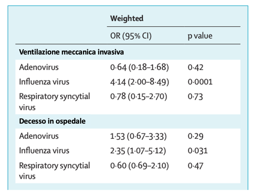
  • Influenza can make you more susceptible to SARS-CoV-2 due to a reduction in physiological defense capabilities. Influenza worsens your symptoms and the risk of hospitalisation. A very recent study conducted in France and published in the Lancet, shows that patients with SARS-CoV-2/Influenza co-infections are more than four times more likely to require assisted ventilation, and 2.35 times more likely to die than patients with SARS-CoV-2 alone. The research, coordinated in the UK as part of the International Severe Acute Respiratory and Emerging Infection Consortium's (ISARIC) – Coronavirus Clinical Characterisation Consortium, is the largest study ever conducted on people with SARS-CoV-2 and other endemic respiratory viruses, with more than 200,000 patients observed.⁹

Figure 2. Use of mechanical ventilation and mortality in the UK from February 2020 to
December 2021 in patients with concomitant SARS-CoV-2 co-infection (OR: Odds Ratio)
  • Co-infection would appear to be a potential risk factor for the exacerbation of lung damage. In a Chinese study, it is shown that in mice that were pre-infected with influenza A, subsequent infection with SARS-COV-2 causes much greater lung damage by the direct effect on ACE2 receptors.¹⁰
  • In general, co-infection of influenza virus and SARS-CoV-2 can cause severe lung infections by other microorganisms for multiple reasons. The influenza virus, for example, increases the infectious potential of opportunistic bacteria, increasing their adherence and invasion ability, making eradicating infections difficult because they require the use of antibiotic therapies and severely debilitate affected patients.¹¹ Modifying the testing strategy for patients with SARS-COV-2 by also expanding the search for influenza much more broadly could therefore be a useful strategy in order to contain other respiratory viral infections and to protect the most fragile population.¹²


The correct and timely identification of Influenza and SARS-COV-2 has practical implications when it comes to managing often similar symptoms and containing the spread of highly transmissible viruses.¹³ The significant increase in risk for patients with co-infection has several implications and suggests that patients hospitalised for SARS-CoV-2 disease should be subjected to dual testing to identify them as potentially more fragile , interpret immunomodulatory responses, or send them for personalised antiviral therapy.


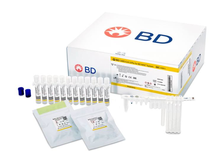
Nasopharyngeal/oropharyngeal swabbing for symptomatic patients can be used to simultaneously detect SARS-CoV-2 Virus, Influenza A and B Viruses, and Respiratory Syncytial Virus-specific genes via the Real Time PCR method. Co-testing for SARS-CoV-2 and other respiratory pathogens such as those indicated using swabs performed on symptomatic patients, is also strongly recommended in the recently published InfluNet 22-23¹⁴ protocol within the context of the Epidemiological and Virology Surveillance by the Supreme Health Council (ISS). As of today, there are highly sensitive molecular tests on the market that can provide a definitive qualitative answer in a short period of time and ensure maximum efficiency of laboratory workflow, such as the BD SARS-Cov-2/Flu test (with the possibility of also detecting RSV in the same reaction) for the BD MAX™diagnostic system.


DOWNLOAD THE BD MAX™ RTI SOLUTIONS BROCHURE


DISCOVER THE BDMAX™ CLUB


BD-73771

Bibliography

1. Ong DSY, Fragkou PC, Schweitzer VA, Chemaly RF, Moschopoulos CD, Skevaki C; European Society of Clinical Microbiology and Infectious Diseases (ESCMID) Study Group for Respiratory Viruses (ESGREV). How to interpret and use COVID-19 serology and immunology tests. Clin Microbiol Infect. 2021 Jul;27(7):981-986.
2. Schulman A. et al, Not Like the Flu, Not Like Car Crashes, Not Like – The New Atlantis. 2020
3. Chotpitayasunondh T, Fischer TK, Heraud JM, Hurt AC, Monto AS, Osterhaus A, Shu Y, Tam JS. Influenza and COVID-19: What does co-existence mean? Influenza Other Respir Viruses. 2021 May;15(3):407-412.
4. Prevalenza e distribuzione delle varianti di SARS-CoV-2 di interesse per la sanità pubblica in Italia. Epicentro ISS. Rapporto 10 dicembre 2021
5. InfluNet & CovidNet. Sistema di Sorveglianza Sentinella delle sindromi simil- influenzali, dei virus influenzali e del virus Sars-CoV-2
6. McKimm-Breschkin JL, Hay AJ, Cao B, Cox RJ, Dunning J, Moen AC, Olson D, Pizzorno A, Hayden FG. COVID-19, Influenza and RSV: Surveillance-informed prevention and treatment – Meeting report from an isirv-WHO virtual conference. Antiviral Res. 2022 Jan;197:105227.
7. Swets MC, Russell CD, Harrison EM, Docherty AB, Lone N, Girvan M, Hardwick HE; ISARIC4C Investigators, Visser LG, Openshaw PJM, Groeneveld GH, Semple MG, Baillie JK. SARS-CoV-2 co-infection with influenza viruses, respiratory syncytial virus, or adenoviruses. Lancet. 2022 Apr 16;399(10334):1463-1464.
8. Stamm P, Sagoschen I, Weise K, Plachter B, Münzel T, Gori T, Vosseler M. Influenza and RSV incidence during COVID-19 pandemic-an observational study from in-hospital point-of-care testing. Med Microbiol Immunol. 2021 Dec;210(5-6):277-282.
9. Swets MC, Russell CD, Harrison EM, Docherty AB, Lone N, Girvan M, Hardwick HE; ISARIC4C Investigators, Visser LG, Openshaw PJM, Groeneveld GH, Semple MG, Baillie JK. SARS-CoV-2 co-infection with influenza viruses, respiratory syncytial virus, or adenoviruses. Lancet. 2022 Apr 16;399(10334):1463-1464.
10. Bai L, Zhao Y, Dong J, Liang S, Guo M, Liu X, Wang X, Huang Z, Sun X, Zhang Z, Dong L, Liu Q, Zheng Y, Niu D, Xiang M, Song K, Ye J, Zheng W, Tang Z, Tang M, Zhou Y, Shen C, Dai M, Zhou L, Chen Y, Yan H, Lan K, Xu K. Coinfection with influenza A virus enhances SARS-CoV-2 infectivity. Cell Res. 2021 Apr;31(4):395-403.
11. Musher DM. Bacterial Coinfection in COVID-19 and Influenza Pneumonia. Am J Respir Crit Care Med. 2021;204(5):498-500. doi:10.1164/rccm.202106-1467ED
12. Saul H, Gursul D, Antonelli M, Steves C. Frail older people and those in deprived areas remain at risk from covid-19, even after vaccination. BMJ. 2022 Jul 4;378:o1313.
13. Pormohammad A, Ghorbani S, Khatami A, Razizadeh MH, Alborzi E, Zarei M, Idrovo JP, Turner RJ. Comparison of influenza type A and B with COVID-19: A global systematic review and meta-analysis on clinical, laboratory and radiographic findings. Rev Med Virol. 2021 May;31(3):e2179.
14. Operational protocol – InfluNet & RespiVirNet season 2022-23 – https://w3.iss.it/site/RMI/influnet/pagine/Documenti.aspx (Viewed on 17 October 2022)


BACK TO THE NEWS

Molecular screening techniques for multidrug-resistant organisms (MDRO) can reduce mortality rates and the economic burden of antimicrobial resistance (AMR)

Molecular screening techniques for multidrug-resistant organisms (MDRO) can reduce mortality rates and the economic burden of antimicrobial resistance (AMR)

Multidrug-resistant organisms (MDROs) are microorganisms (mainly bacteria) that become resistant due to antibiotic misuse as well as through natural mutations.1 Evidence exists that antimicrobial resistance (AMR) is one of the greatest threats to global health, food security and development in today’s society.1 AMR not only prevents the treatment of infectious diseases, such as pneumonia, but according to the World Health Organization (WHO) it also jeopardises the outcomes of modern medicine, as surgeries become much more dangerous without effective antibiotics.1


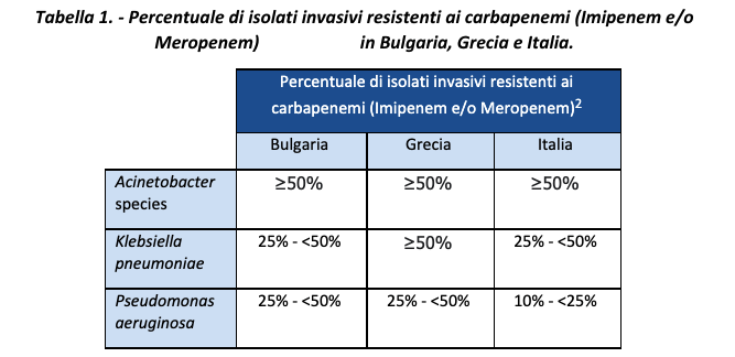
A European surveillance report published in 2020, based on data for 2015-2019, showed that European countries had up to 46.7% of invasive isolates of methicillin-resistant Staphylococcus aureus (MRSA).2 Particularly high rates of resistance to carbapenems were also found, especially in some countries including Greece, Italy and Bulgaria (Table 1).2

Percentage of invasive isolates resistant to carbapenems (Imipenem and/or Meropenem)


Between 2015 and 2019, in various European countries such as Ireland, Germany, Romania, Hungary, Greece, Latvia, Croatia, Lithuania, Poland, and Slovakia, a significant increase was also observed in the Vancomycin-resistant enterococci (VRE) with 25-50% of invasive isolates with resistance.2 Although Norway has one of the lowest rates of AMR in bloodstream infections, resistance to broad-spectrum antibiotics used to combat MDROs has increased in the last10 years.3 Looking specifically at the Italian context, the results of the national AR-ISS surveillance report published at the end of 2021 on the data collected in 2020 at the height of the COVID-19 pandemic still showed high rates of resistance to major antibiotic classes for the 8 pathogens under surveillance, with particular concern for MRSA (it remained stable at around 34%) and Enterococcus faecium (increasing the percentage of vancomycin-resistant isolates to 23.6%).4 


The increase in MDRO prevalence across Europe thus requires effective intervention to reduce the social burden. Several risk factors influence the prevalence of MDROs (some of which emerged recently), which have a big impact on European health systems and communities. This negative impact is likely also due to the shortcomings in current screening techniques, resulting in a lack of detection and control of antimicrobial resistance. 


Risk factors for MDROs in Europe 

The best known risk factor for antimicrobial resistance is “antibiotic misuse”; globally, there is a large amount of antibiotic self-medication due to over-the-counter access. According to the European Centre for Disease Prevention (ECDC) and the WHO, Greece showed a higher defined daily dose (DDD) of antibiotic use in Europe and a 2.4% increase in consumption between 2014 and 2018, while many European countries saw a decrease.4 This, coupled with the high levels of antibiotic resistance in Greece, suggests a causal link.2 


On the other hand, Italy (a country with high levels of antibiotic resistance) showed a relatively low DDD, suggesting the involvement of additional factors. New strains of MRSA were detected in Europe in 2021; in particular, clone t304/ST6 was identified in five northern European countries as originating from the Middle East.6 Moreover, a study of asylum seekers and refugees admitted to Helsinki University Hospital between 2010 and 2017 found that 45% were colonised by MDRO and 12.5% had two or more detectable MDRO strains.7 The percentage of MDRO carriers was highest amongst patients from Iraq (57.2%), Syria (55.8%), Afghanistan (34.8%) and Somalia (25.8%).7 Mass migration from low- and middle-income countries in the Middle East and Africa is prevalent in today's society, with Europe receiving 9.2 million asylum applications between 2010 and 2019.8 According to the European Commission, Italy and Greece are among the European countries with the highest number of first-time asylum applications.9  


A recent study also found an association between MRSA and climate change, suggesting that a 1°C increase in average temperature could result in a 1.02-fold increase.10 There appears to be an association between average warm season temperature and MRSA detection, with a correlation coefficient of 0.826.10 Furthermore, it is estimated that the prevalence of carbapenemase-resistant carbapenemase-resistant Pseudomonas aeruginosa (CRPA) will double in the United Kingdom and the Netherlands by 2039 and increase by 70% in Denmark due to climate change alone.11 Another study in the United States found that a 10°C increase in temperature between regions caused an increase in antibiotic resistance of 4.2%, 2.2% and 2.7% for Escherichia coli, Klebsiella pneumoniae and Staphylococcus aureus; this increase was also associated with higher population density, suggesting that the growth of antimicrobial resistance should not be underestimated in currently growing populations.11    


The impact of AMR on European countries and planned enforcement actions in Italy. 

Lack of sufficient intervention to combat MDROs could lead to a 72% increase in second-line antibiotic resistance by 2030 in the EU/EEA, and last-line antibiotic resistance is likely to double.12 This has serious implications for European countries as infections lead to longer hospital stays, higher rates of antimicrobial resistance, higher medical costs, and increased mortality.13 


Where first-line antibiotics can no longer be used, more expensive drugs are often required, placing a greater economic burden on families and society as a whole.1 In 2019, AMR and its ineffective treatment cost EU/EEA healthcare systems €1.1 billion.12 Alarmingly, in 2019, deaths in Europe due to AMR increased to 33,000.12 A rapid risk assessment for carbapenemase-resistant Enterobacteriaceae (CRE) in 2018 suggested that timely and appropriate laboratory investigations and reporting are essential to avoid delays in appropriate treatment and minimise the associated risk of death.15  


In Italy, the 2022-2025 New National Antibiotic Resistance Plan, drafted by the Ministry of Health and recently published16, is structured around three main areas of action which involve a multidisciplinary and One Health approach (main element of innovation compared to previous plans): 


  • integrated surveillance and monitoring of AMR, antibiotic use, healthcare-associated infections (HAIs) and environmental monitoring;  
  • preventing HAIs in hospital and community settings and infectious diseases and zoonoses;  
  • appropriately using antibiotics in both human and veterinary settings and properly handling and disposing of antibiotics and contaminated materials.


Amongst the objectives in terms of AMR surveillance/monitoring are, for example, strengthening CRE surveillance, promoting the use of molecular methods. The importance of surveilling MDROs nationally had long been under consideration and had been reacknowledged as a necessity even by the AMCLI working group GLISTer in an article in 2017, with a focus on assisted living residences and assimilated local facilities, whilst also identifying three different levels of possible surveillance.


Molecular techniques as a way to supplement to classical cultural techniques 

Traditionally, screening for antimicrobial resistance uses plate culture techniques, where the process of obtaining the result (TAT) from start to finish can take up to five days.18 Waiting this long for identification can lead to delays in implementing appropriate infection control measures, potentially leading to further transmission and mutationi. Delayed identification can also lead to delays in initiating the right treatment, which can also contribute to bacterial resistance and could be detrimental to the patient’s health or even prove fatal.  


One study observed that when specifically comparing the detection of methicillin-sensitive S. aureus (MSSA) and MRSA using culture techniques versus a PCR test, detection is consistently underestimated. For culture methods, direct seeding detected 22.4% of MSSA and 1.2% of MRSA in the samples compared with 35.6% of MSSA and 2.3% of MRSA using a PCR assay.19 In the same study, they modelled these results on national surveillance data and estimated that about 5,000 to 8,000 surgical site infections of S. aureus could be prevented using molecular screening methods. In the UK alone, this could save between £17 million and £130 million per year in treatment costs.19 


There are several issues (and their implications) in the research and development of an effective screening test for MDRO. Molecular diagnostics for MDRO can be useful as a second step to confirm phenotypic culture tests and in surveillance, for example, to confirm the mechanisms responsible for certain resistance20. However, culture methods are believed to heavily underestimate the number of MDROs within a sample, and such false negatives must be minimised to reduce the impact on healthcare systems. This suggests a use of molecular tests as a first step, but although they provide important and clinically relevant information, they themselves have limitations: in fact, molecular tests are more sensitive than culture tests, but they detect only known resistance genes or mutations, and phenotypic resistance testing (culture test) is always necessary in surveillance in the event of a positive result to ensure the correct classification of bacterial isolates20. In any case, it emerged that molecular and phenotypic diagnostics of MDROs complement each other to improve understanding of both the extent of resistance in a given context and the underlying mechanisms responsible for resistance20.  


BD’s commitment and solutions 

BD stands daily alongside healthcare practitioners around the world in the fight against AMR. BD is committed to helping slow the spread of antibiotic resistance by improving awareness, surveillance, infection prevention and patient management. 

We offer a wide range of solutions that can be used to prevent the spread of infections in healthcare facilities, diagnostic systems for monitoring and diagnosing healthcare-associated infections (HAIs), including drug-resistant strains, and state-of-the-art surveillance and reporting capabilities to monitor and predict MDRO outbreaks. 


More specifically: 

  • the range of validated molecular tests for the fully automated BD MAX™ system for monitoring HAIs and major MDROs (including MDR-TB) ensures early and accurate detection which, when combined with the right antimicrobial treatment, can prevent transmission and improve patient management. Learn more about the molecular tests available for CPO, MRSA, VRE and C. diff. [Link to the HAI Molecular Biology brochure/BD Academy information material] 
  • solutions for cultural and phenotypic analysis (manual and electronic), including the BD Phoenix™ M50 and related panels, associated with the BD Bruker MALDI BioTyper® and the BD Epicenter™ middleware which ensures accurate ID/AST results for correct microorganism classification and antibiotic prescription. Find out more HERE.  

BD is an all-round partner in the MDRO surveillance workflow. Learn more about BD’s commitment to combating AMR and HAIs HERE.   


BD-77376

Bibliography

  1. World Health Organization. Antibiotic Resistance. 2020. Available at: https://www.who.int/news-room/fact-sheets/detail/antibiotic-resistance. Accessed: July 2021. 
  2. European Centre for Disease Prevention and Control. Surveillance report. Antimicrobial resistance in the EU/EEA (EARS-Net). 2019. Available at: https://www.ecdc.europa.eu/en/publications-data/surveillance-antimicrobial-resistance-europe-2019. Accessed: July 2021.  
  3. European Centre for Disease Prevention and Control. ECDC country visit to Norway to discuss antimicrobial resistance issues. ECDC. 2019. Available at: https://www.ecdc.europa.eu/sites/default/files/documents/antimicrobial-resistance-country-visit-norway.pdf. Accessed: July 2021.  
  4. Epicenter – AR-ISS Surveillance System Report. Available at: https://www.epicentro.iss.it/antibiotico-resistenza/ar-iss/RIS-1_2021.pdf   – Accessed: Oct 2022. 
  5. Robertson J, Vlahović-Palčevski V, Iwamoto K, Högberg LD, Godman B, Monnet DL, Garner S, Weist K, Strauss R, Vandael E, and Ivanov IN. Variations in the Consumption of Antimicrobial Medicines in the European Region, 2014–2018: Findings and Implications from ESAC-Net and WHO Europe. Front Pharmacol. 2021;12:639207. 
  6. Bartels MD, Worning P, Andersen LP, Bes M, Enger H, Ås CG, Hansen TA, Holzknecht BJ, Larssen KW, Laurent F, and Mäkitalo B. Repeated introduction and spread of the MRSA clone t304/ST6 in northern Europe. Clin Microbiol Infect. 2021;27(2):284.e1-284.e4. 
  7. Aro T and Kantele A. High rates of meticillin-resistant Staphylococcus aureus among asylum seekers and refugees admitted to Helsinki University Hospital, 2010 to 2017. Euro Surveill. 2018;23(45):1700797. 
  8. United Nations High Commissioner for Refugees. Global trends: Forced Displacement in 2019. 2019. Available at: https://www.unhcr.org/5ee200e37.pdf. Accessed: July 2021.  
  9. European Commission. Overall figures of immigrants in European society. 2020. Available at: https://ec.europa.eu/info/strategy/priorities-2019-2024/promoting-our-european-way-life/statistics-migration-europe_en. Accessed: July 2021. 
  10. Kaba HE, Kuhlmann E, and Scheithauer S. Thinking outside the box: association of antimicrobial resistance with climate warming in Europe–a 30 country observational study. Int J Hyg Environ Health. 2020;223(1):151-158. 
  11. MacFadden DR, McGough SF, Fisman D, Santillana M, and Brownstein JS. Antibiotic resistance increases with local temperature. Nat Clim Change. 2018;8(6):510-514. 
  12. European Centre for Disease Prevention and Control. and Organisation for Economic Cooperation and Development. Antimicrobial Resistance: Tackling the Burden in the European Union. 2019; pp.3. Available at: https://www.oecd.org/health/health-systems/AMR-Tackling-the-Burden-in-the-EU-OECD-ECDC-Briefing-Note-2019.pdf. Accessed: July 2021. 
  13. Váradi L, Luo JL, Hibbs DE, Perry JD, Anderson RJ, Orenga S, and Groundwater PW. Methods for the detection and identification of pathogenic bacteria: past, present, and future. Chem Soc Rev. 2017;46(16):4818-4832. 
  14. European Centre for Disease Prevention and Control and the European Medicines Agency. The bacterial challenge: time to react. A call to narrow the gap between multidrug-resistant bacteria in the EU and the development of new antibacterial agents. Available at: https://www.ecdc.europa.eu/sites/portal/files/media/en/publications/Publications/0909_TER_The_Bacterial_Challenge_Time_to_React.pdf. Accessed: July 2021.  
  15. European Centre for Disease Prevention and Control. Rapid Risk Assessment: Carbapenem-resistant Enterobacteriaceae – first update. 2018. Available at: https://www.ecdc.europa.eu/sites/default/files/documents/RRA-Enterobacteriaceae-Carbapenems-European-Union-countries.pdf. Accessed: July 2021.  
  16. Aboutpharma drafting. Antibioticoresistenza: il nuovo Piano nazionale 2022-25 è all’esame delle Regioni. 21 September 2022. https://www.aboutpharma.com/animal-health/antibioticoresistenza-il-nuovo-piano-nazionale-2022-25-e-allesame-delle-regioni/  
  17. Aschbacher, R., Pagani, L., Migliavacca, R. et al. Recommendations for the surveillance of multidrug-resistant bacteria in Italian long-term care facilities by the GLISTer working group of the Italian Association of Clinical Microbiologists (AMCLI). Antimicrob Resist Infect Control 9, 106 (2020). https://doi.org/10.1186/s13756-020-00771-0 
  18. Scerbo MH, Kaplan HB, Dua A, Litwin DB, Ambrose CG, Moore LJ, Murray CCK, Wade CE, and Holcomb JB. Beyond blood culture and gram stain analysis: a review of molecular techniques for the early detection of bacteremia in surgical patients. Surg Infect (Larchmt). 2016;17(3):294-302. 
  19. Tsang STJ, McHugh MP, Guerendiain D, Gwynne P, Boyd J, Walsh TS, Laurenson IF, Templeton KE, and Simpson AHRW. Underestimation of Staphylococcus aureus carriage associated with standard culturing techniques. Bone Joint Res. 2018;7(1):79-84. 
  20. Global Antimicrobial Resistance Surveillance System (GLASS). Molecular methods for antimicrobial resistance (AMR) diagnostics to enhance the Global Antimicrobial Resistance Surveillance System. Geneva: World Health Organization; 2019 (WHO/WSI/AMR/2019.1). Licence: CC BY-NC-SA 3.0 IGO. https://www.who.int/publications/i/item/WHO-WSI-AMR-2019.1  


BACK TO THE NEWS

Vaginal swabs with a self-sampling procedure for HPV DNA testing in the prevention of cervical cancer

Vaginal swabs with a self-sampling procedure for HPV DNA testing in the prevention of cervical cancer

In 2020 the World Health Organization (WHO) launched a plan of action to achieve the elimination of cervical cancer caused by Human Papilloma Virus (HPV) through the vaccination and treatment of at least 90% of women by 2030¹.

Much had already been done before the pandemic, through awareness campaigns on the fundamental issue of prevention¹-².


Italy has locally organised screening programmes, which for decades have provided free access to cervical screening/HPV DNA testing for women of 25 to 64 years of age, bringing the incidence of cancer to lower levels than the world average. However, the incidence is not yet at zero, and it is therefore important to maintain high levels of vigilance and to continually improve access and adherence to preventive tests³. As attested by the IARC since 1996, HPV infection is the essential condition for the development of cervical cancer: it is the first cancer to be recognised by the World Health Organisation as being totally attributable to infection. This was discovered in 1976 by Prof. Harald Zur Hausen, who was awarded the Nobel Prize 30 years later, in 2008.


There are approximately 120 types of HPV viruses, but only 12 cause cervical cancer. Testing positive for HPV does not necessarily mean you have cancer at that time, but it does mean you are at greater risk of it developing in the future. Most of these infections resolve spontaneously with no symptoms, but an infection that persists for more than two years can result in precancerous lesions and carcinoma⁴. In 2020, cervical cancer was the fifth most common cancer in women in Italy under 50 years of age(2,400 new cases estimated in 2020, accounting for 1.3% of all cancers affecting the female population⁵.


Eliminating cervical cancer is now a global public health goal launched by the WHO in 2018, and a commitment of the European Union, which has included it in Europe's Beating Cancer Plan⁶. The aim of screening for cervical carcinoma is to detect persistent infections caused by types of viruses that have oncogenic potential, and para-neoplastic lesions, in order to implement appropriate follow-up and treatment before an actual tumour develops³.


Nowadays, it is no longer necessary to see a gynaecologist for HPV testing. Patients can also choose to collect a sample themselves at home and take it to a licensed diagnostic centre, where they will get a clear result quickly in the interests of staying healthy. All under the supervision of a practitioner who will manage follow-up for the woman in the event of a positive result. HPV DNA testing is simple and effective, and women can also collect samples independently thanks to self-sampling procedures using suitable validated⁷-⁸ devices. With self-sampling, which is already practised when screening to prevent colorectal cancer for example, women can collect a sample of cells for HPV DNA testing themselves at home, without having to go to a clinic or gynaecologist, making it even easier to participate in the screening campaign. A number of clinical studies⁹ have been carried out to assess the reliability and accuracy of the results obtained by self-sampling, and both women and practitioners can trust the procedure.


To screen for HPV infection, Becton Dickinson offers BD Onclarity™ HPV test, which is performed in the laboratory on BD Viper™ LT (for small- to medium-sized laboratories) and BD COR™ (for high productivity centralised laboratories) automated platforms. The test fully complies with the European Regulation, being CE IVD marked, and is clinically validated according to the Meijer¹⁰ criteria for applicability in cervical screening using the primary HPV DNA test. The test is CE-IVD validated on specimens collected to additionally perform liquid cytology (cervical screening test) in BD SurePath™ or ThinPrep® vials.


BD Onclarity™ HPV test makes it possible to:

detect the DNA of the 14 HPV genotypes that carry oncogenic risk (HR- HPV16, 18, 31, 33, 35, 39, 45, 51, 52, 56, 58, 59, 66, and 68) associated with the development of cervical cancer, with the possibility of extended genotyping in addition to HPV genotypes 16 and 18. Six genotypes are genotyped individually, and the remaining are genotyped into 3 different groups (P1, P2 and P3)

analyse the genes for viral oncoproteins E6 and E7, reducing the risk of false negative results that can be found with DNA analysis in the L1 gene, following the integration of viral DNA into cellular DNA

reduce false positive results due to cross-reactivity with HPV genotypes with low oncogenic risk

ensure the detection of possible co-infection by the 14 different genotypes with high oncogenic risk


MUSA™ – Be inspired by simplicity

Out of these assumptions, and the desire of BD to become a pioneer and at the forefront working alongside Italian laboratories and women in supporting women's health and screening against HPV, MUSA™, an initiative created to raise awareness of the importance of cervical cancer prevention through the diagnosis of Human Papillomavirus, has emerged.


In close collaboration with the Italian Diagnostic Center – CDI, MUSA™ promotes the importance of screening as an indispensable weapon with which to fight this disease, thanks also to the innovative HPV Test in self-sampling mode: convenient, easy and effective, which can be used wherever and whenever the patient wishes. By filling in a simple request form, patients can purchase the self-sampling kit, which will be delivered to their home. This enables them to collect a sample for the HPV test independently and send it to the Laboratory by post/courier. This method of sampling was designed to try to maximise and facilitate participation in screening programmes for women who find it difficult or embarrassing to attend a clinic¹¹-¹².


In recent years, clinical studies conducted both in Italy and internationally have sought to determine both the satisfaction of self-collection and the safety and efficacy of the HPV DNA test performed on self-collected samples compared with those collected by a practitioner (gynaecologist/obstetrician). The results confirmed thereliability of self-sampling and a high level of acceptance among women ¹¹-¹².


Do you want to find out more? Click on the banner below and follow MUSA™ on Instagram and Facebook.


BD-83680

References


  1. Global strategy to accelerate the elimination of cervical cancer as a public health probmem. OMS 2020
  2. European guidelines for quality assurance in cervical cancer screening. 2015
  3. Camara H, Zhang Y, Lafferty L, Vallely AJ, Guy R, Kelly-Hanku A. Self-collection for HPV-based cervical screening: a qualitative evidence meta-synthesis. BMC Public Health. 2021 Aug 4;21(1):1503.
  4. Gisci website. https://www.gisci.it/il-nuovo-programma-di-screening-con-il-test-hpv-sostituisce-il-pap-test. Visited 27 February 2022
  5. Redazione ANSA. https://www.ansa.it/canale_saluteebenessere/notizie/sanita/2022/05/31/hpv-in-italia-quinto-tumore-per-le-donne-under50_364100ce-cd7c-4410-9ca2-e4190785e0da.html Visitato il 01 giugno 2022
  6. Europe’s Beating Cancer Plan
  7. Rossi P, Marsili LM, Camilloni L, Iossa A, Lattanzi A, Sani C, Di Pierro C, Grazzini G, Angeloni C, Capparucci P, Pellegrini A, Schiboni ML, Sperati A, Confortini M, Bellanova C, D’Addetta A, Mania E, Visioli CB, Sereno E, Carozzi F. The effect of self-sampled HPV testing on participation to cervical canrcer screening in Italy: a randomised controlled trial (ISRCTN96071600). British Journal of Cancer (2011) 104, 248 – 254.
  8. Snijders P, Verhoef V, Arbyn M, Ogilvie G, Minozzi S, Banzi R, van Kemenade F, Heideman D, Meijer C. High-risk HPV testing on self-sampled versus clinician-collected specimens: A review on the clinical accuracy and impact on population attendance in cervical cancer screening. International Journal of Cancer (2013) 132(10):2223-36
  9. Canfell K, Smith MA, Bateson DJ. Self-collection for HPV screening: a game changer in the elimination of cervical cancer. Med J Aust. 2021 Oct 18;215(8):347-348.
  10. BD Onclarity™ HPV Assay EU Package Insert (8089899).
  11. Del Mistro A, et al. Efficacy of self-sampling in promoting participation to cervical cancer screening also in subsequent round. Prev Med Rep . 2016 Dec 23;5:166-168. doi: 10.1016/j.pmedr.2016.12.017. eCollection 2017 Mar
  12. Arbyn M et al. Detecting cervical precancer and reaching underscreened women by using HPV testing on self samples: updated meta-analyses. BMJ. 2018 Dec 5;363:k4823. doi: 10.1136/bmj.k4823.


BACK TO THE NEWS